

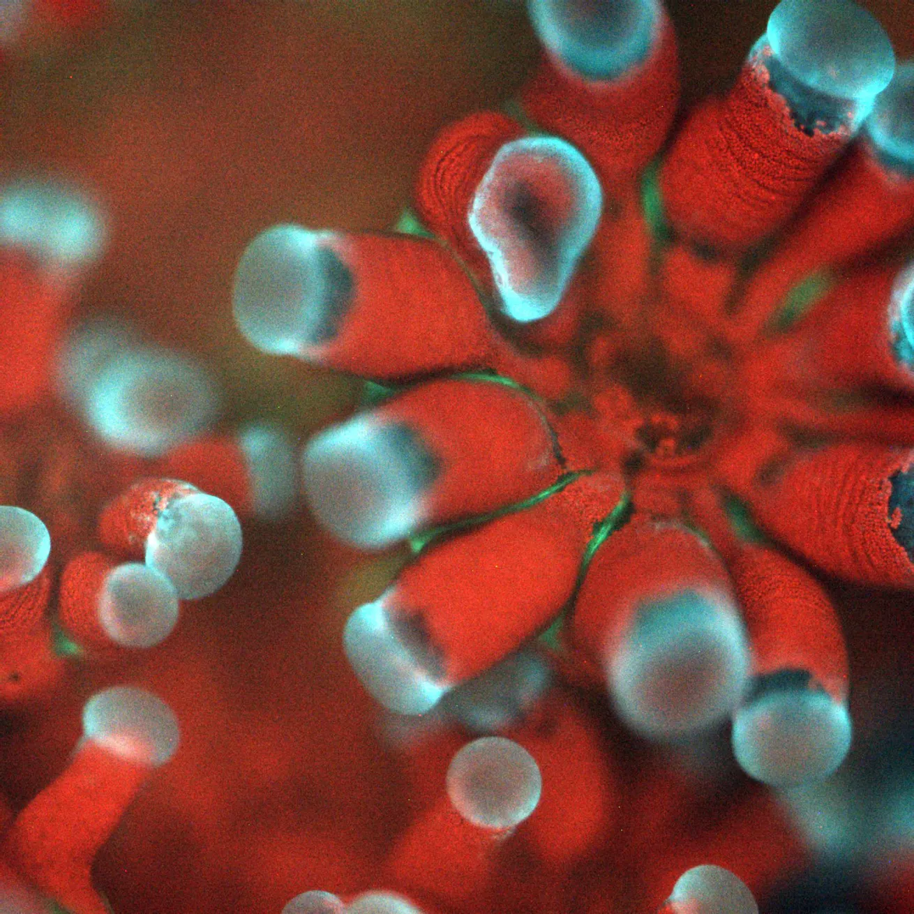
Microscope Photo by Or Ben-ZviAn image of a Stylophora pistillata coral taken with the BUMP. Each polyp has a mouth and a set of tentacles, and the red dots are individual microalgae residing inside the coral tissue.
A newly developed diver-operated microscope promises to reveal the secrets of coral bleaching by bringing the relationship between corals and their symbiotic algae into focus in an innovative way.
Known as BUMP (the Benthic Underwater Microscope imaging PAM), the device combines two types of technology—underwater microscopy and lab-based pulse amplitude modulated (PAM) light techniques. Underwater microscopy allows researchers to observe the microalgae living inside corals, while PAM measures photosynthesis. A paper about the microscope was recently published in the journal Methods in Ecology and Evolution.
“By combining these two [technologies], we got a very powerful tool that gives us a lot of structural but also physiological information about the coral’s health,” says lead author Or Ben-Zvi, a postdoctoral researcher at the University of California, San Diego’s Scripps Institution of Oceanography.
Corals survive thanks to a symbiotic partnership with tiny algae, called zooxanthellae, which live inside the coral’s tissue. These brightly colored algae give corals their vivid hues and provide them with food.
Related Reading: Finding Family-Friendly Diving in Fiji
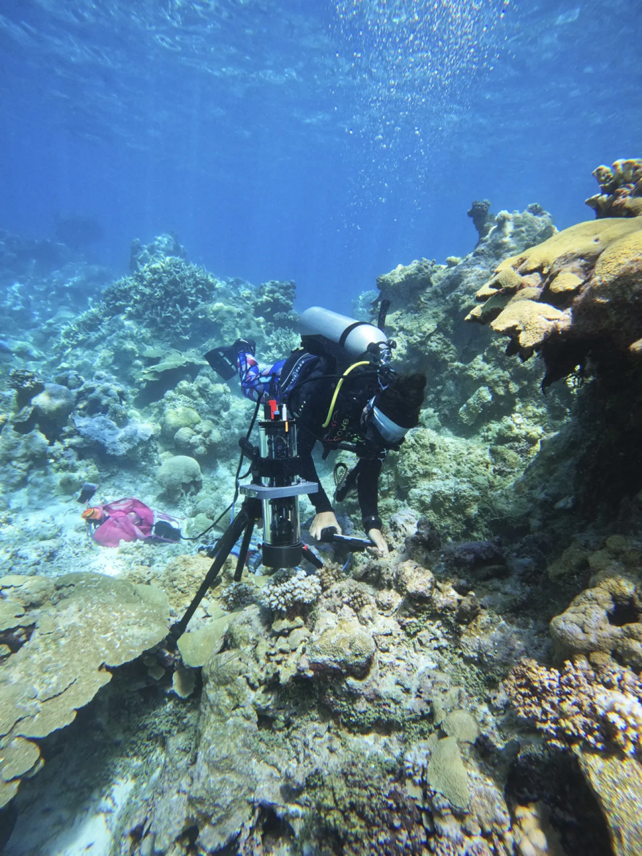
Photo by Samantha Clements, Courtesy or Ben-ZviA field deployment of the BUMP in Palmyra Atoll, where corals were imaged and measured.
Corals can filter organic particles from the water, but they get most of their energy from the zooxanthellae. While living inside the coral, the algae use photosynthesis to convert sunlight into energy and share this with their host—almost like a little thank you for letting them stay.
But this relationship is very delicate. If the conditions change—particularly if the water gets too hot—corals can become stressed and expel their colorful zooxanthellae, leaving the coral to appear bleached white.
Although bleached coral isn’t dead, it is more vulnerable to disease and can quickly starve without its key food source. If the conditions return to normal and the microalgae can come back before too long, the coral can recover. But scientists still have more to learn about exactly what happens when corals bleach. That’s where the BUMP microscope comes in.
“We assume that prior to the stage of coral bleaching… there are changes in their photosynthetic efficiencies,” Ben-Zvi says. The BUMP can see and measure photosynthesis at the scale of individual microalgae within the coral, so scientists hope it could spot the warning signs that a bleaching event might be on the way.
Related Reading: Marine Biologist Jacinta Shackleton Named April '25 Sea Hero
“We hope that we can detect these early signs of stress, before the microalgae are expelled and the coral bleaches,” she says. “If we are able to detect the underlying mechanisms or can provide early alerts in specific areas, we might be able to prevent or at least alleviate some of the pressure the corals are under.”
Ben-Zvi was surprised by how much the microscope could reveal about an area of coral measuring just 2 square millimeters—even within this tiny space, the algae’s ability to photosynthesize changed quite significantly.
“As a marine biologist who is used to watching the corals as big colonies with less detail, it was amazing to see their micro life,” she says. “They are more detailed and more active than I imagined.”


